Research area
Our Vision
The Nanotrio refers to a class of diverse nanomaterials developed at the intersection of nanophotonics, nanoplasmonics, and nanobiotechnology. Our research explores the synthesis, characterization, and potential applications of nanomaterials, with a particular focus on quantum dots, plasmonic nanoparticles, and nanocomposites with tunable morphologies and sophisticated nanostructures.
We specialize in characterizing the “small world” of nanomaterials using absorption and emission spectroscopy, transmission electron microscopy, dynamic light scattering, and zeta-potential analysis. Through these analyses, we investigate the properties of synthesized materials and use the resulting insights to guide the future design of functional nanomaterials.
Our goal is to understand the unique physical properties of nanomaterials and translate them into practical applications in nanotherapy, nanoenergy, nanodiagnostics, and other emerging fields.
Ongoing Research Projects (For more information, please visit the Project page)
Our Nanotrio laboratory conducts research in nanoscience and nanobiology, focusing on the interaction between light and matter at the nanoscale. We study the synthesis, surface control, assembly, and complexation of nanocrystals to develop nanostructures and advanced materials with potential applications in displays, photodetectors, photovoltaics, photoelectrochemical cells, sensors, and bioimaging. In particular, we study semiconductor nanocrystals, such as quantum dots and QLED materials, as well as nanostructured materials based on gold nanoparticles that can be used for sensing and three-dimensional cell imaging. By leveraging the unique physical properties of nanomaterials, we aim to develop functional materials for a wide range of optical, electronic, and biomedical applications.
We synthesize new nanocrystals by controlling precursor reactivity and reaction intermediates. Quantum dots are inorganic nanocrystals with a molecular-level size distribution, and their small size allows a large number of atoms to reside at the surface. For example, InP quantum dots, which are widely used in QLED displays, can contain approximately 1,000 to 10,000 atoms, depending on the size. Although the exact atomic structure and growth mechanism of these nanocrystals remain difficult to determine, quantized intermediates can form during their synthesis. These intermediates are structurally analogous to the critical nuclei described in classical nucleation theory. Our research focuses on understanding these intermediate nanoclusters and controlling their formation through precursor chemistry, with the goal of establishing growth pathways and mechanistic principles for the development of high-performance QLED nanocrystals.
We also investigate surface chemistry at the nanoscale. Surface chemistry is essential for forming efficient conjugates with proteins, DNA, and antibodies, and it plays a critical role in controlling colloidal stability and surface functionality. In particular, we study ultrafast surface modification using quantized ions and investigate reversible surface bonding based on hydrogen-bonding interactions. Through these approaches, we aim to establish precise and efficient strategies for tailoring nanocrystal surfaces.
Another major focus of our research is near-infrared optical materials. Compared with conventional molecular or organic materials, quantum dots can emit in the near-infrared region while exhibiting high photostability and size-tunable optical properties. Quantum-dot-based optical probes can also be miniaturized and used for ultrahigh-sensitivity sensing through solution-based processes such as inkjet printing. Using these properties, we develop new near-infrared fluorescent probes and materials. In addition, we are developing brain–computer interfaces that use autonomous quantum-dot probes to visualize real-time physiological signals in the brain, thereby enabling new approaches for understanding human cognition and neural activity. We are also synthesizing biocompatible nanocrystals and converting them into oil-in-fluorocarbon materials for biomedical applications.
We develop nanorobotic probes that respond to environmental stimuli, such as pH, by changing their optical properties. Like cuttlefish that change color in response to their surroundings, these nanorobotic probes are designed to alter their optical signals when exposed to specific environments. Quantum-dot optical probes can also be used as next-generation display materials because they provide a wide color gamut and can respond sensitively to external stimuli. By programming their response to specific conditions, these probes can visualize changes that are difficult to detect directly. For example, quantum dots that respond to magnetic field variations are being investigated as magnetoplasmonic nanorobot probes that use gold nanoparticles and can be applied to next-generation transparent displays, including AR and VR technologies.
Finally, we are developing barcode technologies using near-infrared probes to overcome the limitations of conventional molecular probes and to enable simultaneous detection of multiple targets. Because of their strong photostability, high brightness, and narrow emission bandwidth, quantum dots can provide highly multiplexed optical signals. Antibody–quantum dot conjugates can be used for rapid and quantitative target detection in complex biological environments. These nanomaterials offer advantages for medical diagnosis, bioimaging, and precision sensing by minimizing biological background interference and providing distinct multicolor optical signals. Through this research, we aim to develop quantum-dot-based molecular barcode platforms capable of detecting multiple professional-level diagnostic signals simultaneously.
진행 중인 연구 분야
● 나노트리오 연구실은 나노스케일에서 빛과 물질의 상호작용을 이해하고 제어하는 것을 목표로, 나노과학 및 나노바이오 분야의 연구를 수행하고 있습니다. 본 연구실은 나노결정의 합성, 표면 제어, 조립 및 복합화 연구를 통해 디스플레이, 광검출기, 태양전지, 광전기화학 소자, 센서, 바이오이미징 등 다양한 분야에 활용될 수 있는 나노구조체와 첨단 소재를 개발하고 있습니다. 특히 양자점 및 QLED 소재로 활용되는 반도체 나노결정과, 센싱 및 3차원 세포 이미징에 적용 가능한 금 나노입자 기반 나노구조체를 중점적으로 연구하고 있습니다. 이를 통해 나노소재 고유의 물리적 특성을 활용하여 광학, 전자, 바이오의학 분야에서 기능성 소재로 발전시키는 것을 목표로 합니다.
● 본 연구실은 전구체의 반응성과 반응 중간체를 제어함으로써 새로운 나노결정을 합성하는 연구를 수행하고 있습니다. 양자점은 분자 수준의 크기 분포를 갖는 무기 나노결정으로, 매우 작은 크기 때문에 많은 수의 원자가 표면에 존재합니다. 예를 들어 QLED 디스플레이에 널리 사용되는 InP 양자점은 크기에 따라 약 1,000개에서 10,000개의 원자로 구성될 수 있습니다. 이러한 나노결정은 정확한 원자 구조와 성장 메커니즘을 규명하기 어렵지만, 합성 과정에서 양자화된 중간체가 형성될 수 있습니다. 이러한 중간체는 고전적 핵생성 이론에서 설명하는 임계핵과 구조적으로 유사한 나노클러스터로 볼 수 있습니다. 본 연구실은 이러한 중간 나노클러스터의 형성과 반응성을 이해하고, 전구체 화학을 통해 이를 제어함으로써 고성능 QLED 나노결정 개발을 위한 성장 경로와 성장 원리를 확립하고자 합니다.
● 또한 나노스케일에서의 표면화학을 연구하고 있습니다. 표면화학은 단백질, DNA, 항체 등과의 효율적인 결합체 형성에 필수적이며, 콜로이드 안정성과 표면 기능성을 조절하는 데 중요한 역할을 합니다. 특히 본 연구실은 양자화된 이온을 이용한 초고속 표면 개질과 수소결합 상호작용을 기반으로 한 가역적 표면 결합을 연구하고 있습니다. 이러한 접근을 통해 나노결정 표면을 정밀하고 효율적으로 조절할 수 있는 전략을 확립하고자 합니다.
● 다른 주요 연구 주제는 근적외선 광학 소재입니다. 일반적인 분자 또는 유기 소재와 비교할 때, 양자점은 높은 광안정성과 크기 조절이 가능한 광학 특성을 가지면서 근적외선 영역에서 발광할 수 있습니다. 또한 양자점 기반 광학 프로브는 소형화가 가능하고, 잉크젯 프린팅과 같은 용액 공정을 통해 초고감도 센싱 플랫폼으로 활용될 수 있습니다. 본 연구실은 이러한 특성을 활용하여 새로운 근적외선 형광 프로브와 소재를 개발하고 있습니다. 더 나아가, 자율적으로 작동하는 양자점 프로브를 통해 뇌의 생리적 신호를 실시간으로 시각화하고, 이를 기반으로 인간의 인지 및 신경 활동을 이해하기 위한 뇌-컴퓨터 인터페이스 기술을 개발하고 있습니다. 또한 생체적합성 나노결정을 합성하고 이를 오일-인-플루오로카본 소재로 전환하여 바이오의학 응용으로 확장하는 연구도 수행하고 있습니다.
● pH와 같은 환경 자극에 반응하여 광학 특성이 변화하는 나노로봇 프로브를 개발하고 있습니다. 주변 환경에 따라 색을 변화시키는 갑오징어처럼, 이러한 나노로봇 프로브는 특정 환경에 노출되었을 때 광학 신호가 변화하도록 설계됩니다. 양자점 기반 광학 프로브는 넓은 색 영역을 구현할 수 있고 외부 자극에 민감하게 반응할 수 있어 차세대 디스플레이 소재로도 활용될 수 있습니다. 특정 조건에 반응하도록 프로그래밍된 프로브는 직접 관찰하기 어려운 변화를 시각화할 수 있습니다. 예를 들어 자기장 변화에 반응하는 양자점은 금 나노입자를 이용한 자기-플라즈몬 나노로봇 프로브로 연구되고 있으며, AR/VR 기술과 같은 차세대 투명 디스플레이 응용으로 확장될 수 있습니다.
● 마지막으로, 기존 분자 프로브의 한계를 극복하고 여러 표적을 동시에 검출하기 위해 근적외선 프로브 기반 바코드 기술을 개발하고 있습니다. 양자점은 높은 광안정성, 강한 발광 세기, 좁은 발광 대역폭을 가지므로 다중화된 광학 신호를 제공할 수 있습니다. 항체–양자점 접합체는 복잡한 생물학적 환경에서도 표적을 빠르고 정량적으로 검출하는 데 활용될 수 있습니다. 이러한 나노소재는 생체 배경 신호의 간섭을 줄이고, 서로 구분 가능한 다색 광학 신호를 제공함으로써 의료 진단, 바이오이미징, 정밀 센싱 분야에서 장점을 가집니다. 본 연구실은 이러한 연구를 통해 여러 진단 신호를 동시에 검출할 수 있는 양자점 기반 분자 바코드 플랫폼을 개발하고자 합니다.
Nanophotonics
The emission wavelength and intensity of QDs can be precisely controlled, making them versatile materials for switchable probes in cellular and whole-body imaging, optical modulation, and light-emitting devices. Our group synthesizes a wide range of nanomaterials, including core/shell QDs, nanorods, nanotetrapods, and nanoporous structures. We have demonstrated the photomodulation of QD conjugates using additional UV excitation, which promotes electron transfer to the QDs and quenches their photoluminescence. We have also developed inorganic-ligand-capped QDs as sensitizers for solar-cell applications.
1. Optical properties of quantum dots
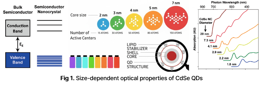
Semiconductor nanocrystals, commonly referred to as quantum dots (QDs), differ from bulk semiconductors in that their optical properties are strongly dependent on size. In semiconductor nanocrystals with diameters of only a few nanometers, electrons and holes are confined within a space smaller than the exciton Bohr radius, giving rise to quantum confinement in all three dimensions. This confinement produces discrete electronic energy levels and makes the band gap (BG) between the valence band and the conduction band dependent on nanocrystal size. The BG determines the energy required for photoexcitation and the subsequent radiative relaxation process. As the size of a QD decreases, the BG increases, leading to absorption and fluorescence at shorter wavelengths (Fig. 1). QD core/shell structure can be classified into type-I and type-II (Fig. 2)
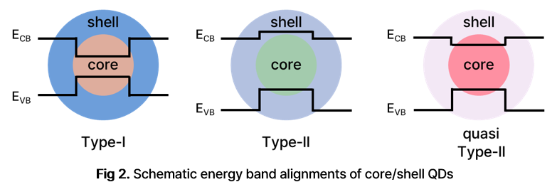
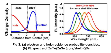
Semiconductor QDs can be categorized into type-I and type-II structures according to their band alignment. In type-I QDs, the shell material has a larger BG than the core material, allowing the electron–hole pair to be confined within the core. This confinement enhances photoluminescence (PL) efficiency, making type-I QDs useful for highly emissive applications. In type-II QDs, the valence and conduction band edges of the core and shell are staggered, which spatially separates electrons and holes across the core/shell interface (Fig. 3a). This charge separation makes type-II QDs attractive for energy-conversion applications, including solar cells. Changes in the valence and conduction band alignment of the core and shell alter the spatial distribution of electron and hole densities. This band engineering also creates an effective BG that is not determined solely by the intrinsic band gaps of the constituent materials, enabling control over both absorption and PL wavelengths (Fig. 3).
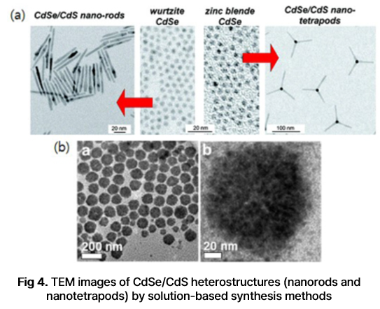
Colloidal nanocrystals synthesized from solution-phase precursors offer broad tunability in morphology and size, providing many opportunities for functional applications. Morphological control is important because nanocrystal shape and internal structure influence electronic energy states, charge-carrier mobility, and exciton localization. These morphologies can be controlled by regulating nucleation and growth processes from the atomic level. Such processes are governed by both kinetic factors, including reactant concentration and diffusion, and thermodynamic factors, including surfactant concentration and reaction temperature. Solution-based synthesis can produce highly crystalline nanocrystals with diverse shapes and offers notable advantages over conventional top-down methods. We synthesize various types of nanocrystals, including core/shell QDs, nanorods, nanotetrapods, and nanoporous materials (Fig. 4a). We have also reported supra-QDs (SQDs) (Fig. 4b), which are hierarchical superstructures composed of hundreds of individual QDs, each only a few nanometers in size, assembled through oriented attachment. These SQDs may exhibit enhanced charge-carrier mobility and therefore show strong potential as sensitizers for solar-cell applications.
2. Investigation of the properties of NPs induced by the incorporation of foreign atoms
Introducing foreign atoms into as-synthesized nanocrystals can alter their optical, electrical, and magnetic properties in addition to changing their composition. Depending on the amount and distribution of the introduced species, this process can be classified in several ways. One representative approach is doping, in which semiconductor nanocrystals are modified in a manner analogous to the doping of bulk semiconductors.
Doping is the intentional incorporation of foreign atoms into a pristine semiconductor host material to modulate its optical, electrical, and magnetic properties. In bulk semiconductors, dopants ionize and provide additional electrons or holes depending on their valence states. In the case of transition-metal dopants, the dopant spin can couple with the orbitals of the host material. Because of these characteristics, doped semiconductors are widely used in semiconductor junction structures, including transistors and diodes.
In semiconductor nanocrystals, however, doping is strongly affected by quantum confinement and therefore produces behaviors distinct from those observed in bulk materials. The confinement of excitons within an extremely small volume enhances their interaction with dopant ions, amplifying changes in optoelectronic and magnetic properties. Doped nanocrystals therefore have potential applications in memory devices, photocatalysts, and bioimaging probes, in addition to conventional semiconductor junction structures.
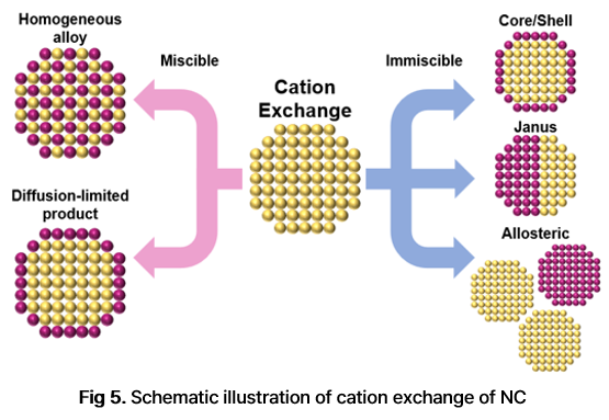
Cation exchange is another method for introducing foreign atoms into a crystal lattice. Because bonding in many semiconductor nanocrystals has substantial ionic character, newly introduced cations can replace original cations while preserving the anion framework. This transformation enables the synthesis of sophisticated nanostructures, including metastable crystal phases and complex morphologies that are difficult to access through direct synthesis.
By controlling the amount of foreign ions, cation exchange can be used for doping, heterostructure formation, or the synthesis of entirely new materials. The exchange reaction is driven by differences in the thermodynamic stability of the reactants and products, as well as by ligand-mediated complexation under specific reaction conditions. The feasibility of a given exchange reaction can be evaluated by considering the reaction conditions, solubility products, and standard enthalpies of formation of the relevant materials (Fig 5).
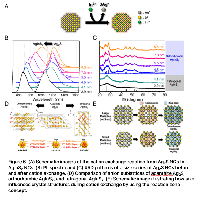
In nanocrystals, even when the constituent materials are identical, the crystal structures obtained after cation exchange can differ depending on particle size because thermodynamic stability is size-dependent (Fig. 6). Therefore, investigating how cation-exchange outcomes vary with nanocrystal size is important for understanding the reaction mechanism. After cation exchange, optical spectroscopy, X-ray diffraction, energy-dispersive X-ray spectroscopy, and other atomic-level characterization methods should be used to confirm the composition and crystal structure of the final material.
3. PL modulation of QD
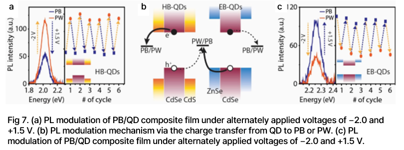
The emission wavelength and intensity of QDs can be modulated, making them promising materials for switchable probes in cellular and whole-body imaging, optical modulation, and light-emitting devices. We used quasi type-II QDs to develop photoluminescence-modulating probes that respond to external stimuli. Charging quasi type-II QDs, in which electrons and holes are spatially separated, induces photoluminescence (PL) spectral switching. The initial emission state can be recovered through a subsequent neutralization step. Reversible PL switching over many repeated cycles was demonstrated by alternating UV excitation between on and off states (Fig. 7). We also developed a modulation sensor based on a PL on/off system. In addition, reversible and wavelength-selective PL modulation was investigated by controlling charge transfer between QDs and quenchers.
4. QD-based Solar Cells
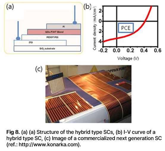
A solar cell (SC) converts light energy directly into electrical energy. Although silicon SCs have been commercialized on a large scale, they remain costly and require complex fabrication processes. Nanocrystal-based SCs offer a potentially simpler and more cost-effective alternative. Several types of SCs use QDs, including QD-sensitized SCs, hybrid SCs composed of QDs and conducting polymers (Fig. 8), and Schottky-type SCs.
Although these SCs have different device architectures, they all rely on light absorption followed by charge generation, separation, and transport. Charge separation and transfer are influenced by the morphology of the QDs and the structure of the active layer. The use of QDs allows the absorption wavelength to be tuned, and their colloidal dispersibility in various solvents enables solution-processable device fabrication. Therefore, QD-based SCs represent a promising research direction for developing low-cost solar-energy conversion technologies. By identifying the conditions required for high power conversion efficiency, experimental studies can provide insights into simple and scalable fabrication strategies for QD-based SCs.
Nanoplasmonics
Surface plasmon resonance (SPR) arises when free electrons in metal nanoparticles collectively oscillate in resonance with incident electromagnetic energy at the interface between the metal and the surrounding dielectric medium. Noble metal nanoparticles exhibit strong SPR when their dimensions are reduced to the nanoscale, and their optical properties can be precisely tuned by controlling the metal composition, particle size and shape, and local dielectric environment. These tunable SPR characteristics make noble metal nanoparticles highly versatile platforms for spectroscopic analysis, optical imaging, sensing, and therapeutic applications. Our group develops advanced metal nanoparticle complexes for highly efficient cancer therapy and photoacoustic-guided drug delivery.
- Properties of Surface Plasmon Resonance
Conductive metals contain a high density of free electrons that are not strongly localized to individual atoms. Because these electrons can collectively respond to external electromagnetic fields, metallic nanostructures can exhibit surface plasmon resonance (SPR) when their dimensions are reduced to the nanoscale. SPR gives metal nanoparticles unique optical properties. In particular, noble metal nanoparticles, such as gold and silver nanoparticles, interact strongly with visible light and exhibit intense absorption and scattering, with optical cross sections that are orders of magnitude larger than those of typical organic dyes. They also show high resistance to oxidation and photobleaching, making them powerful optical probes that can complement or replace conventional organic fluorophores.
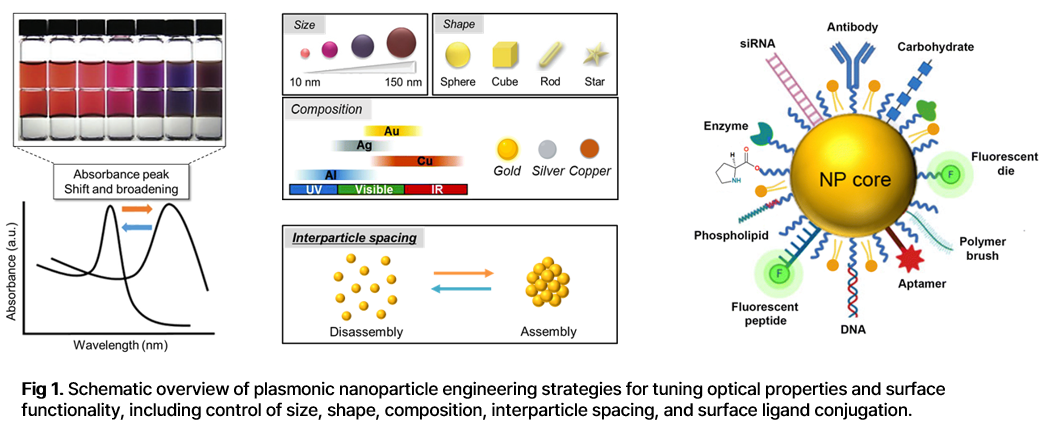
The optical responses of noble metal nanoparticles are highly tunable because their plasmon resonance can be controlled by the dielectric properties of the metal, as well as by the size, shape, and local environment of the nanostructure. Our goal is to design and synthesize diverse metallic nanostructures and to investigate their optical properties using spectroscopic and imaging tools. We further aim to apply these nanostructures to biological and biomedical systems (Fig. 1).
- Study of Thermotherapy for Tumor Cells
Photothermal therapy is a cancer treatment strategy in which absorbed photon energy is converted into heat to induce cancer cell death (Fig. 2). Because cancer cells are generally more sensitive to elevated temperatures than normal cells, selective heat generation within tumor tissues can improve therapeutic efficacy while reducing damage to surrounding healthy tissue. Metal nanoparticles are highly effective photothermal agents because of their strong light absorption and efficient photothermal conversion. By controlling the thermal energy generated through the photothermal effect of metal nanoparticles, selective cancer therapy can be achieved.

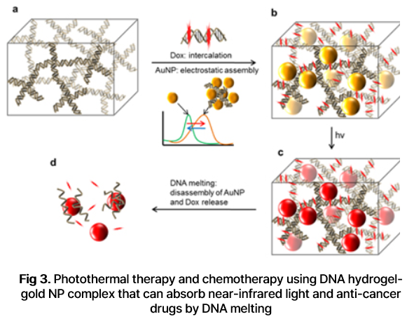
Near-infrared (NIR) light is widely used for photothermal therapy because it penetrates deeply into living tissues and is only weakly absorbed or scattered by major biomolecules such as hemoglobin and water. The therapeutic selectivity of pH-responsive “smart” gold nanoparticles that absorb NIR light preferentially in cancer cells or tumor tissues can be further enhanced by exploiting the acidic and hypoxic microenvironment of tumors. In addition, nanoparticle-mediated photothermal treatment can be combined with anticancer drugs to achieve synergistic therapeutic effects. Gold nanoparticle aggregation can be induced through various strategies. For example, DNA hydrogel–nanoparticle complexes can be designed to release anticancer drugs while simultaneously promoting nanoparticle aggregation, providing a platform for combined photothermal and chemotherapeutic treatment (Fig 3).
- Dark-Field Microscopy
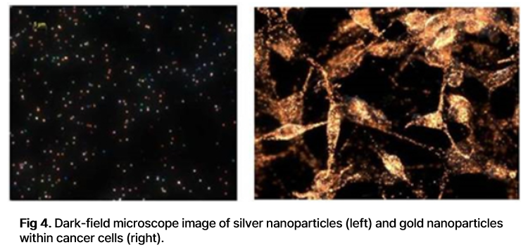
Dark-field microscopy (DFM) visualizes scattered light from a specimen, whereas conventional transmission-mode microscopy forms images using transmitted light. Metal nanoparticles can generate strong contrast under DFM because they efficiently scatter incident light at wavelengths that match their SPR. Because scattering signals from small metal nanoparticles are intense and highly sensitive to plasmonic properties, DFM enables optical visualization even at the single-particle level.
DFM is also useful for distinguishing materials that may show only weak contrast in conventional imaging modes. This capability allows the SPR properties of individual metal nanoparticles to be characterized and enables biological imaging using metal nanoparticles as scattering-based contrast agents (Fig. 4).
- Photoacoustic Imaging Using “Smart” Gold Nanoparticles
Photoacoustic imaging (PAI) is an emerging hybrid imaging modality that combines optical excitation with ultrasound detection. As a nonionizing biomedical imaging method, PAI can provide molecular and functional information, including hemoglobin oxygen saturation, blood flow, and tissue morphology. It is therefore well suited for biomedical studies, including the monitoring of tumor physiology and treatment responses.
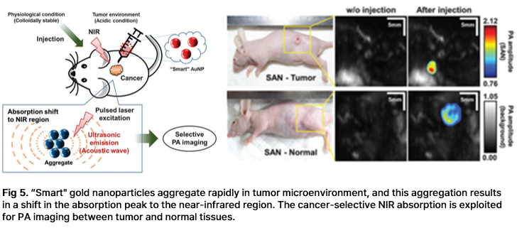
In PAI, image contrast is determined by the optical absorption properties of the target. Exogenous contrast agents with strong near-infrared (NIR) absorption can enhance PA signals because NIR light penetrates deeply into biological tissues. “Smart” gold nanoparticles (SANs) can rapidly aggregate under mildly acidic conditions, which are commonly found in tumor microenvironments. This aggregation induces a red shift in their absorption spectrum toward the red and NIR regions. As a result, PA amplitude increases under mildly acidic conditions while remaining relatively unchanged at physiological pH. By combining tumor accumulation with high PA conversion efficiency, SANs can significantly amplify PA signals in cell-level and small-animal experiments (Fig. 6). These properties make SANs a promising platform for PA-guided drug delivery, enabling real-time identification of delivery targets and on-demand therapeutic administration.
Nanobiotechnology
QD-based optical probes provide a promising platform for in vivo bioimaging and next-generation medical imaging applications because of their bright luminescence, resistance to photobleaching, and size-tunable emission wavelengths. Our group develops NIR-II-emitting QD probes and has demonstrated cancer diagnosis using QD–antibody conjugates. We have also engineered amphiphilic polyethylenimine-derivative platforms that incorporate QDs for cell labeling, targeted delivery, gene delivery, and ratiometric oxygen sensing.
1. Biological applications of Quantum dots
Semiconductor QDs present a possible new technology for in vivo bio-imaging and future medical-imaging applications. QDs have proven potential as imaging contrast agents due to their bright luminescence, their resistance to photobleaching, and their tunable emission wavelengths. The main impediment to the use of QDs for biological applications is their biocompatibility. QDs that contain toxic elements such as Cd, Pb, Hg, or As have limited biological applications. High quality ‘non-toxic’ QDs have been synthesized (e.g., InP, ZnTe/ZnSe, CuInS2; AgInS2); they are free from potential leakage of toxic heavy-metal ions, and photoluminesce at wavelengths that can be selected by adjusting the size of the QD. The new methods of synthesizing ‘non-toxic’ QDs using various solvents and QD precursors have been investigated to obtain bright and photostable QDs that promise application to in vivo medical imaging.
2. Surface modification, functionalization and bioconjugation of QDs for biological applications
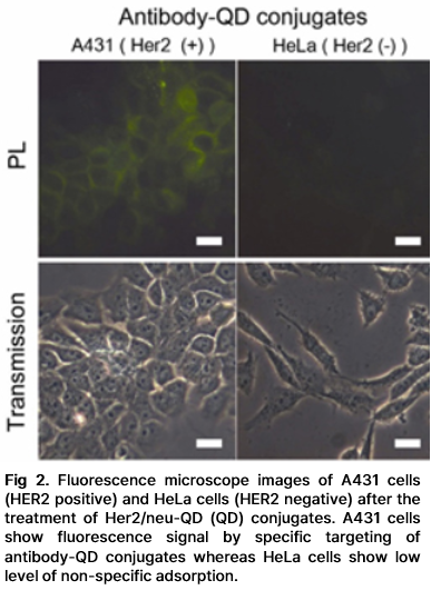
QDs are synthesized in inorganic solvent to obtain high quality, so their surfaces should be modified for bioimaging applications. The surface molecules for QDs should give good colloidal stabilities in aqueous solutions without aggregation, and should have appropriate sites to allow conjugation with biomolecules. To be suitable for applications in targeted imaging or in long-term in vivo imaging, QDs must be colloidally stable and must have minimal non-specific binding. QDs that have been modified to have zwitterionic surface molecules can maintain colloidal stability over broad ranges of pH and ionic strength, and to show minimal non-specific adsorptions (Fig. 1). Moreover, the zwitterionic QD surface ligands can also be used in conjunction with other functional groups; this trait allows simple conjugations for highly specific targeting while retaining the advantages of a zwitterionic QD surface (Fig. 2).

3. Cancer Diagnosis Using QD - Antibody Conjugates
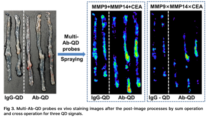
Cancer is a very heterogeneous system. Multiplexed QD imaging is well-suited to detecting such heterogeneous systems. QDs are also bright, photostable, and allow multiplexing by a single excitation wavelength. QDs have broad absorption profile with large one-photon and two-photon absorption cross sections, narrow and symmetric emission profile, and large effective Stokes shifts. To improve the accuracy of cancer detection, multiple biological targets must be imaged concurrently; these advantages of QDs can be used in multiplexing to achieve fluorescence molecular imaging of cancer tissues. Doctors frequently use endoscopy to detect colon cancer, but the diagnosis is based on the clinician's naked-eye perception, which is subjective and error-prone.We developed a rapid and accurate molecular fluorescence imaging technique that uses antibody (Ab)-coated QDs (Ab-QDs) that are sprayed and washed simultaneously on colon tumor tissues inside live animals, then excited and imaged by endoscopy. QDs were conjugated to matrix metalloproteinase (MMP) 9, MMP 14, or carcinoembryonic antigen (CEA) Abs with zwitterionic surface coating to reduce nonspecific bindings. The Ab-QD probes can diagnose tumors on sectioned mouse tissues, in fresh mouse colons stained ex vivo and in vivo, and in fresh human colon adenoma tissues in 30 min and can be imaged to a depth of 100 μm. The probes successfully detected not only cancers that are readily discernible to the naked eye, but also hyperplasia and adenoma regions. Sum and cross-signal operations provided post-processed images that can show complementary information or regions of high priority (Fig. 3). This multiplexed QD, spray-and-wash, and endoscopy approach provides a significant advantage for detecting small or flat tumors that may be missed by conventional endoscopic examinations and bestows a strategy for the improvement of cancer diagnosis.
4. NIR-II emitting QD probes
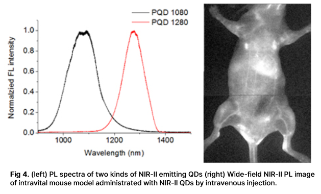
The NIR biological window from 650 nm to 1700 nm allows deeper penetration than visible light into biological tissues such as skin and blood, because these tissues scatter and absorb less light at long wavelengths than at short wavelengths. The window between 650 nm and 950 nm (NIR-I) has been exploited extensively for in vivo optical imaging of live animals, but little research has considered use of the longer-wavelength window (NIR-II) from 1000 to 1700 nm for in vivo fluorescence imaging of live animals. Light scattering and absorption by biomolecules are much weaker in the NIR-II region than in NIR-I, so tissue penetration depth and spatial resolution are increased, and autofluorescence is decreased in NIR-II. QDs can be advantageous fluorescent probes for molecular imaging in both the NIR-I and the NIR-II region. CdTeSe, and CuInS2 have been developed as fluorophores in NIR-I, and have shown high photostability and quantum yield (QY) > 50%. PbS and Ag2S QDs have been developed for NIR-II fluorescence imaging applications, and showed QY > 10% with inorganic shells in the aqueous phase (Fig. 4). We report an enzyme-activatable NIR-II probe that exhibits fluorescence (FL) upon matrix metalloprotease activity in tumor microenvironment. Bright and stable PbS/CdS/ZnS core/shell/shell QDs were synthesized as a model NIR-II fluorophore, and activatable modulators were attached to exploit photoexcited electron transfer (PET) quenching. The quasi-type-II QD band alignment allowed rapid and effective FL modulations with a compact surface ligand modulator that contains methylene blue PET quencher. The modulator was optimized to afford full enzyme accessibility and high activation signal surge when the enzyme was active. In a colon-cancer mouse model, the probe demonstrated selective FL activation at tumor sites with tripled signal strength in 10 min (Fig. 5).
We report an enzyme-activatable NIR-II probe that exhibits fluorescence (FL) upon matrix metalloprotease activity in tumor microenvironment. Bright and stable PbS/CdS/ZnS core/shell/shell QDs were synthesized as a model NIR-II fluorophore, and activatable modulators were attached to exploit photoexcited electron transfer (PET) quenching. The quasi type-II QD band alignment allowed rapid and effective FL modulations with a compact surface ligand modulator that contains methylene blue PET quencher. The modulator was optimized to afford full enzyme accessibility and high activation signal surge when the enzyme was active. Using a colon cancer mouse model, the probe demonstrated selective FL activation at tumor sites with tripled signal strength in 10 min (Fig. 5).

5. Sensing molecule by using QDs
Amphiphilic polyethyleneimine derivatives (amPEIs) were synthesized and used to encapsulate dozens of QDs. The QD-amPEI composite was ~100 nm in hydrodynamic diameter and had an outer surface with a slight positive charge that is well suited for cellular internalization. The outer positive surface was further exploited for gene delivery and targeting. Co-delivery of QDs and GFP-silencing RNAs was demonstrated by attaching siRNAs to the outer surface; the process achieved transfection efficiency that was an order of magnitude higher than conventional gene transfections. Hyaluronic acids were tethered onto the QD-amPEI for cell-specific targeted labeling, which achieved a specific-to-nonspecific signal ratio > 100. The inside hydrophobic compartment was further applied to cohost oxygen-sensing phosphorescence Ru dyes along with QDs. The ratiometric PL signals of the QD-Ru-amPEI oxygen probe show accurate and reversible oxygen-sensing capability and were successfully applied to cellular and spheroid models (Fig. 6).
